
ZEISS VersaXRM 5 Insight accelerates hybrid 3D X-ray imaging
New system combines X-ray microscopy and microCT for seamless multi-scale imaging in one connected workflow
ZEISS today introduced ZEISS VersaXRM 5 Insight, a hybrid 3D X-ray microscope designed to help researchers and engineers understand samples faster. Combining X-ray microscopy and microCT in a single platform, the system delivers multi-scale imaging, guided workflows, and automated AI-enabled reconstruction for efficient non-destructive 3D imaging. Built for performance and engineered for value, ZEISS VersaXRM 5 Insight helps researchers and engineers in materials research, industrial inspection, and life sciences reduce tradeoffs between throughput, resolution, and usability.
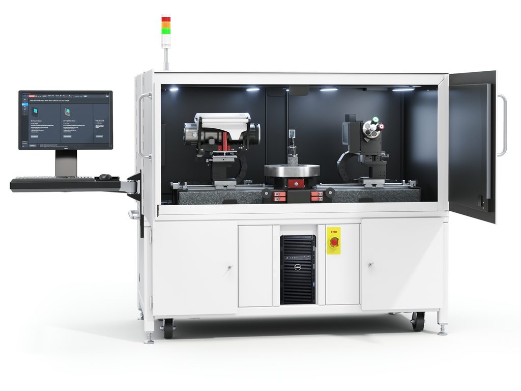
Multi-scale imaging without workflow disruption
Researchers, engineers, and life scientists often need to examine complex samples at different length scales to reveal internal features that affect performance, behavior, or failure, or to understand the structure and spatial organization of biological specimens. ZEISS VersaXRM 5 Insight addresses this need by combining large field-of-view and high-resolution imaging in one system. Users can inspect larger samples, zoom into regions of interest, and capture detailed 3D insights without changing hardware configurations or interrupting their workflow or experiment.
The system combines the throughput, flexibility, and efficiency of microCT with the detailed imaging and insights of lab-based X-ray microscopy. This hybrid capability enables a unified workflow for imaging both large-scale structures and fine internal features while preserving sample integrity.
High-resolution imaging which preserves sample integrity
ZEISS VersaXRM 5 Insight includes Resolution at a Distance (RaaD), which enables high-resolution imaging of small features and internal defects within larger samples. By reducing the need to cut, trim, or refixture samples, the system helps users across science and industry save time, reduce preparation errors, and protect delicate or difficult-to-reproduce specimens.
This non-destructive imaging approach helps users identify regions of interest inside a sample before additional preparation or follow-up analysis. This enables more targeted investigations, reduces unnecessary sample processing, and improves confidence in inspection, research, and quality assurance applications.
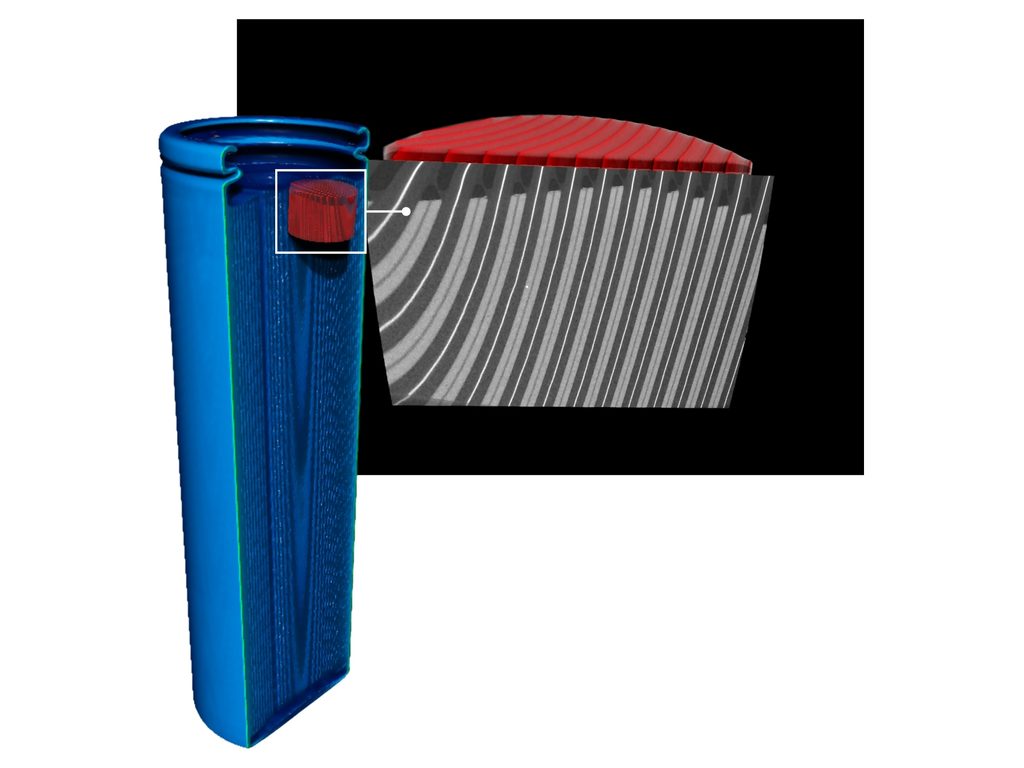
ZEISS VersaXRM 5 Insight flat panel imaging of a cylindrical 21700 battery (70 mm × 21 mm) showing full cell on left using flat panel at 70 µm (60 minutes), and a magnified view of red inset region on the right acquired at high resolution (3.5 µm, ~9.5 hours) highlighting the internal layered structure for detailed, non-destructive characterization.
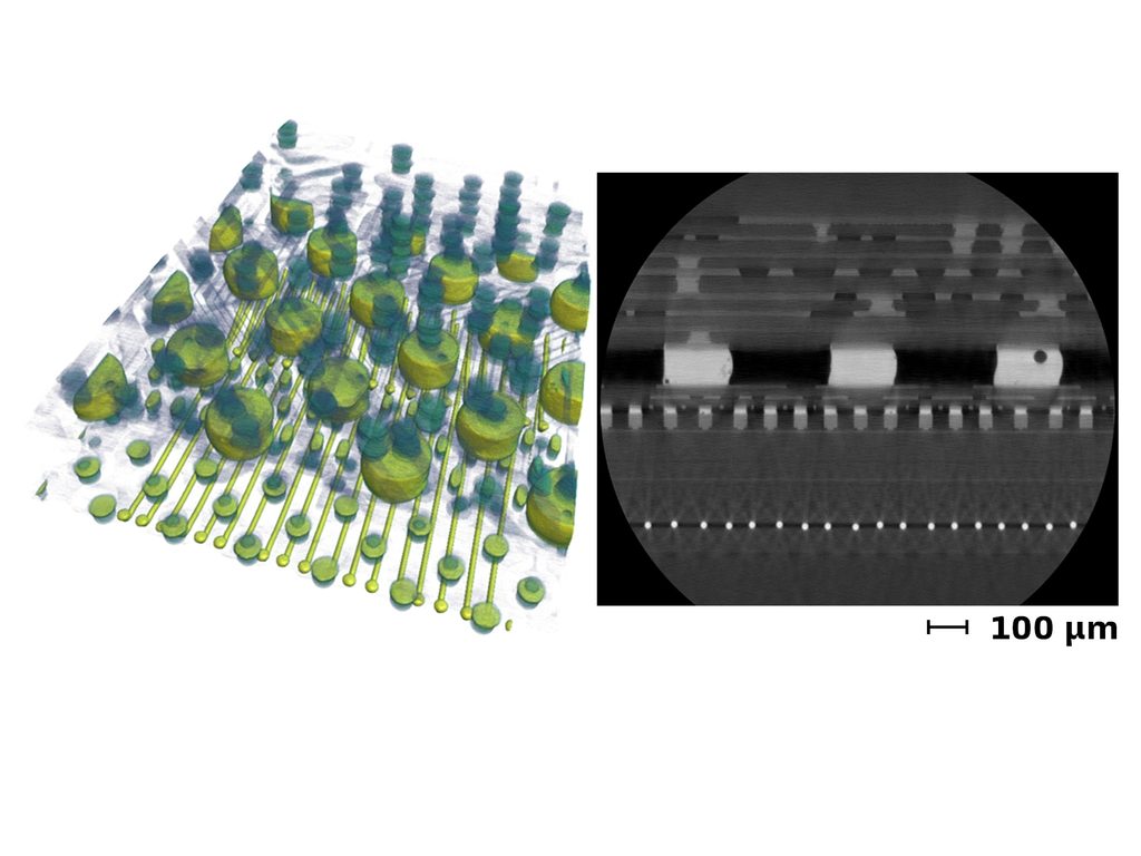
ZEISS3D reconstruction of a 5G modem package on a smartphone board imaged with ZEISS VersaXRM 5 Insight. The left image shows the C4 bump and microbump architecture and the right image displays a reconstructed slice of the corresponding internal interconnect structure acquired at 0.8 μm / voxel using a 4x objective (~1.5 hours).

Dr. Keith Duncan, Danforth Plant Science Center, Missouri, USAArabidopsis thaliana flower fixed in buffered aldehydes, contrasted with osmium tetroxide, embedded in agarose, and imaged with ZEISS VersaXRM 5 Insight using the 4x objective. Datasets processed with ZEISS arivis Pro. Sample courtesy of Dr. Keith Duncan, Danforth Plant Science Center, Missouri, USA.
Guided operation for faster, more reliable results
Productivity in 3D X-ray imaging depends on more than scan speed. ZEISS VersaXRM 5 Insight addresses setup time, operator variability, and workflow decisions with ZEN navx, an intuitive software environment with intelligent guidance and sample-aware navigation. This software simplifies setup, supports sample-specific imaging decisions, and protects both the sample and the system.
FAST Mode adds rapid imaging feedback for three-dimensional navigation, whole-part inspection, and intermediate-resolution scans. This helps users accelerate screening, positioning, and in-situ experiments while improving the overall time to results.
“For teams that need both day-to-day productivity and advanced 3D imaging capability, ZEISS VersaXRM 5 Insight offers a practical path forward,” said Dr. Keith Duncan, Research Scientist and Director of X-ray Imaging at the Danforth Plant Science Center, Missouri, USA, after beta-testing the system. “The improved detector architecture, which combines a flat-panel detector with Resolution at a Distance (RaaD), delivers impressive image quality. It helps users move from overview to detail quickly while keeping samples intact and workflows streamlined.”
Designed for research and industry
ZEISS VersaXRM 5 Insight supports diverse applications in materials research, industrial inspection, and life sciences. In materials research, it helps users investigate failure mechanisms, detect internal defects, and quantify microstructural evolution with in situ and 4D studies. In industrial workflows, it supports high-resolution part quality inspection, root-cause analysis in semiconductor and electronic packages, and yield improvement through deeper 3D insight across manufacturing, quality assurance, and failure analysis.
In life sciences, ZEISS VersaXRM 5 Insight enables researchers to visualize biological structures in 3D across multiple scales, identify regions of interest for follow-up imaging studies, and preserve valuable samples.
“ZEISS VersaXRM 5 Insight combines the flexibility of microCT, the detail of X-ray microscopy, and guided workflows in a single value-optimized platform,” said Nicolas Gueninchault, Head of the X-ray Microscopy Field of Business at ZEISS Microscopy. “With this system, users can work more efficiently, protect valuable samples, and gain the 3D insight they need without being forced to choose between throughput, resolution, and ease of use.”